


Valgus feet deformation is most congenital. But in some cases - paralysis, with traumatic lesions - can no longer be seen during life. The main signs of pathology are the visible violations of the legs and muscles of the legs and muscles of the legs and the muscles of the lower leg, as well as pain in gait change. The diagnosis of the disease is carried out using a clinical examination, X -Ray, electromatography, etc. Treatment includes conservative and surgical methods. At the same time, the right effectiveness is only observed in reconstructive operations.
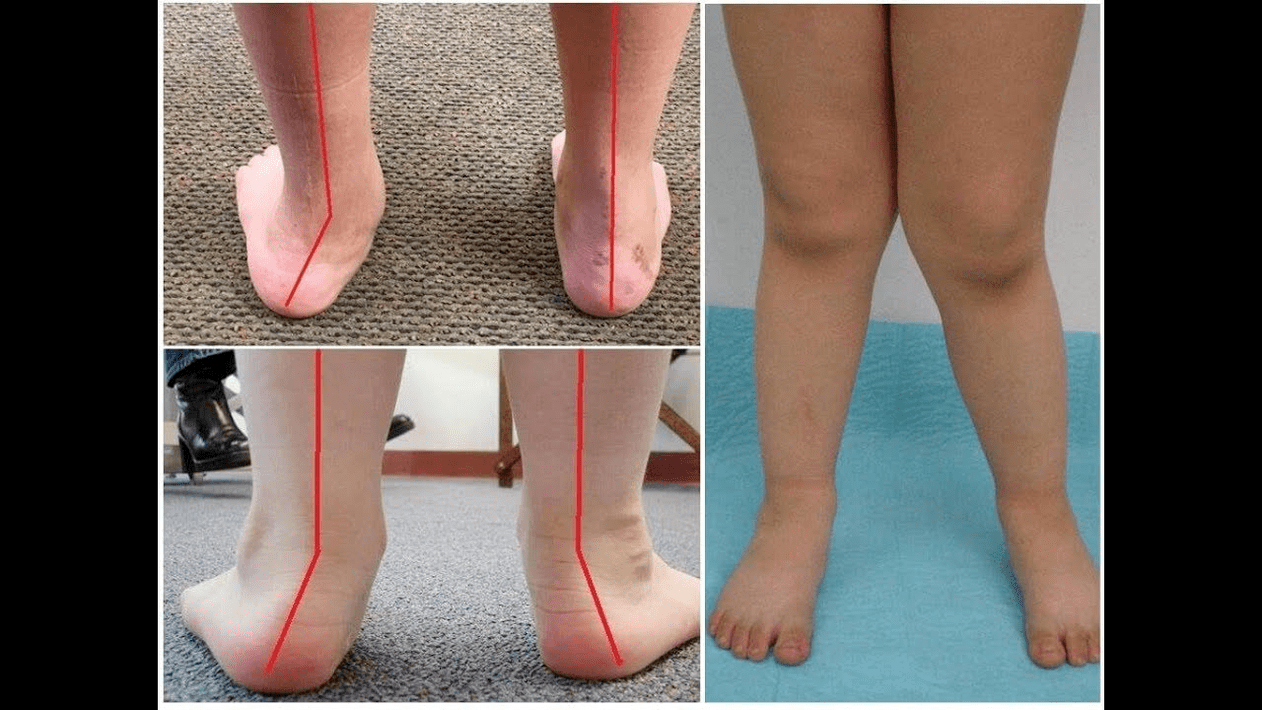
Valgus deformation is the curvature of the foot characterized by the collapse of the longitudinal arch. Usually, the inner edge of the foot ("drops") are reduced and the heel is leaving.
It takes the pressure of a person's leg, the place of the human body, respectively. For this reason, it has a special anatomical structure that allows depreciation, balancing and stabilization. However, an important component for the implementation of these tasks is the correct form of stops.
Today, the most important problem of traumatology and orthopedics is the deformation of the skirt. The meeting is estimated at 30-58%, here are congenital disorders in 2/3.
The pathological, mainly socially important, because the population covers all age groups and helps early development of the spinal column, the joints of the jets of the lower extremities and early development of arterrais.
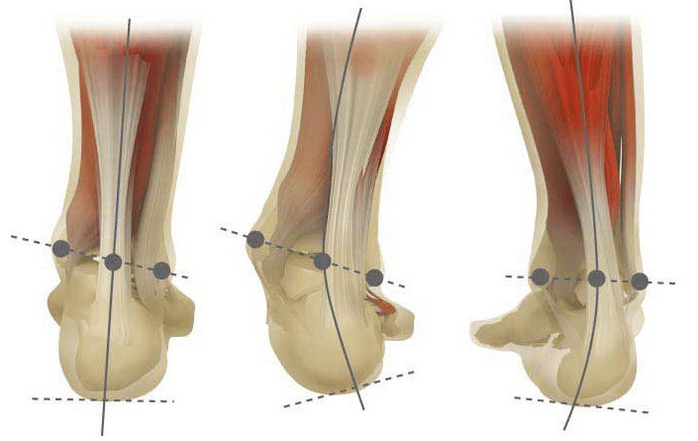
When you pick up the legs (if you look at them from behind), a deformation occurs at an ankle level: the ankle is in contact, the heels are 5-6 centimeters.
Most are congenital in pathological nature and diagnosed in the hospital (or immediately after starting the march). A similar situation is adjusted to 5 years, after which one child develops straight legs.
The main reason for the main cause of the foot of the leg is the weakness of the posterior tibial muscle or the weakness of the Ligamanabi apparatus.
Today, other factors in the development of pathology are different:
- The wrong location of the bones of the legs or shortening of tendons (vertical RAM bone, short pile tendon);
- Test violations while compensating the curvature of the deformation of the legs;
- Traumatic lesions (rupture of the bones, lower legs, hips or knees, ligament and tendon);
- Encephalitis, polyomyelite, stroke, because of damage to the nervous system for violation of sterebrospinal, paralysis (immobilization).
- Spasm of the muscles of the lower leg (constant contraction);
- Sever diseases: vitamin D (Rickets), diabetes Mellitus, Osteoporosis (reduction of bone density), impaired thyroid and paratiroid glands, etc. ;
- In menopause or pregnancy, body weight, including rapid weight loss, has increased.
The development of pathology is also facilitated by the extreme correction of the wrong selected shoes or the clubfote.
The severity of pathology (power of manifestation) is divided into degrees:
- 1, 5-2 centimeters and light light with a sloping angle to 15 degrees;
- When the ARC is flattened to the 1st centimeter and the angle decreases to 10 degrees;
- The angle of 0, 5 cm and heel in the height of the tonga is 5 degrees.
Depending on the participation of certain structures, the following stages of the curvature differ:
- There are no bone deformities, the pain is determined on the internal surface of ankle (in the fastener area of posterior tibes);
- The curvature is light, the heel is slightly rejected;
- The foot separates and deformation is fixed (not corrected correctly);
- Sustainable not only on foot, but also observed in the ankles attachment.
In the first stage, patients are worried about periodic pain after walking or long vertical loads (sitting on the feet). As a rule, pain syndrome is strengthening while walking in the properly selected shoes. The next stage of the disease is associated with the occurrence of the curvature of the foot: patients in a constant position do not trust the outside edge of the foot, but with the whole area. A little change is observed on the ground.
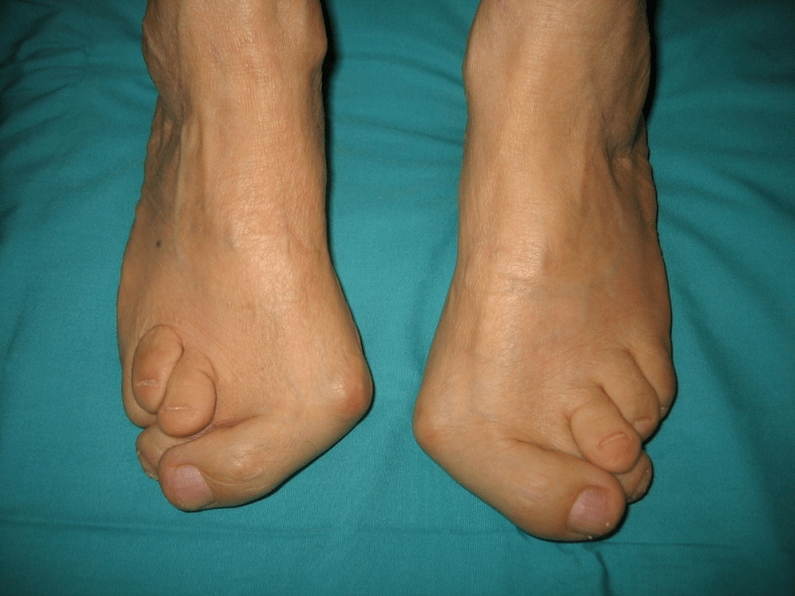
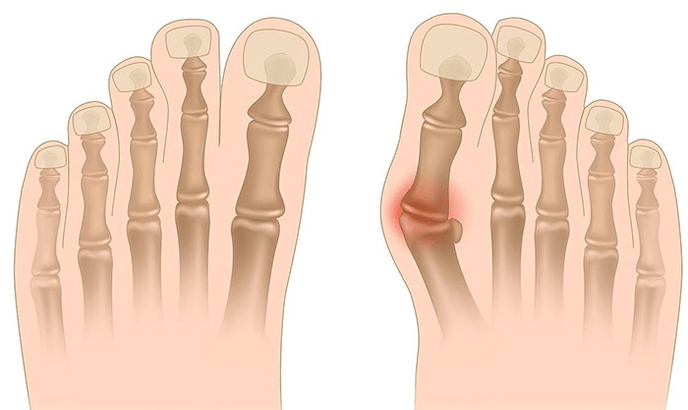
In the third stage, the protrusions of the melting bone are determined (less than ankle on the inside surface of the ankle), as well as a strong distraction of the heel (based on the inner edge of the patient's heel bone). The advanced valgus deformation of the legs is characterized by the curvature of the legs of the legs and the ankle joint. Patients complain of severe pain in the lower foot muscles, as well as an important gait violation: knees are located slightly, right and left foot.
The heavy bend of the legs is often complicated by deformation of the spine (different positions of the shoulders), osteocondrosis or arthrosis) or arthrosis (premature wear of the arthrosis).
The diagnosis of the curvature of the foot consists of:
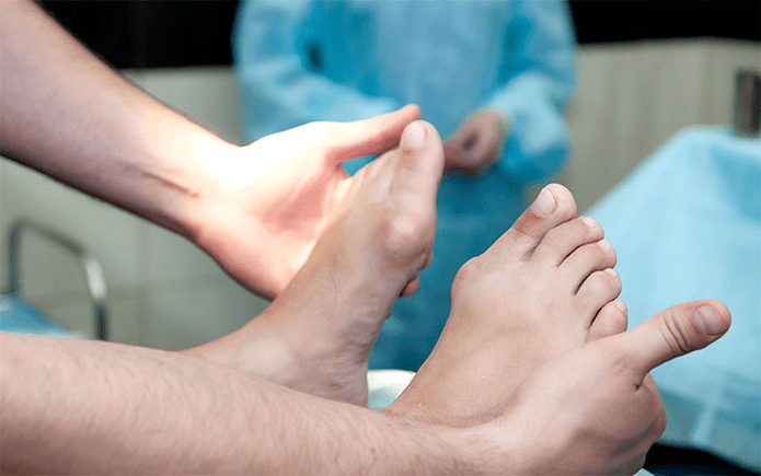
- Clinical inspection reveals that the deviation of the arches of orthopedists, foot arches, heels and rams, the deviation of the arched "disappearance".
- X -ray - a suitable and informative method that you can set changes in the linear settings of changes and connections in the bone tendency angles. These indicators need to make the latest diagnosis and clarify the deformation rate.
- The method of registration of notes in order to determine the accurate functional status of the mayor. The method is to mark the time of the support of the individual parts of the foot when taking a step. During the study, the foot rolling phases are also studied by the balance of the muscles of the lower extremity.
- Dynamic electromyography that marks the dependence of the studied muscles and the dependence on the step stage.
- Photoplomography with digital processing that allows you to access all standard indicators and determine the type / degree of the type of curvature.
An additional consultation of a neurologist (deformations due to spasms or paralysis), endocrinologist (in the case of diabetes / thyroid / parathyroid gland diseases or disorders) and the gynecologist (in case of fear). If the curvature of the foot appears in the background of osteoporosis, densitometry is necessary - the study of bone density.
Among the main methods of treatment of valgus curvature, conservative and operation is carried out. Do not destroy painful joints with ointment and injection!
Conservative approach
This help is aimed at getting rid of the symptoms of the disease, but does not overcome the root cause of pathology.
The technique includes:
- I Plus the use of orthopedic insoles to eliminate the back of the bone, the corner of the foot, as well as the middle of the leg and the valgus lesions;
- Taping - to correct your leg and ankle using special glue ribbons with proper elasticity. Band tape is worn over the hours for 3-5 days, then they are replaced;
- build orthopedic shoes according to individual standards;
- The use of oroz and other adjustment devices in foot and ankles.
Conservative methods also include physiotherapeutic procedures (ozokerite, paraffin applications, electrophoresez, magnetic effects), massage and physiotherapy training complex designed for a specific clinical work. Be careful! Today, most specialists prefer treatment methods because conservative therapy is ineffective (according to statistics, 60% of the work is useless).
Surgical intervention
The volume of the transaction and its type depends on the direct stage of the disease. Thus, the first degree of Valgus deformity is treated by the heel (split) to return to the synonymeal (the extraction of tendon crust to correction) or anatomically. In the second stage of the development of the disease, the transplantation of the tendon bends of the fingers is used. Such an intervention is carried out against the background of the division of the heel or Ram-lobbed arthrodis (the surgical immobilization of the compound between the bones of ram and the scaphid).
The curvature of the III is at a time requires an increase in several plays of the foot: an increase, five cups of cubic and RAM. This kind of triple immobilization is often completed by the divination of the heel bone. In the 4th phase of pathology, reconstructive operations are not only on foot, but also in ankle. In this case, the non-stability of the ligamentaous apparatus is regulated using the transportation of (from its own body or artificial materials). The volume of transactions in the foot is the same as the degree of curvature of the III.
Recovery period
Rehabilitation includes a walk without supporting the operated foot within 2 months. At the same time, the patient should wear a gypsum that can be removed from 1, 5 to 3 months. After the operation, active actions are recommended to start 1, 5 months after the operation. In the 3th month, a physical education complex is applied. However, afterwards, the patients are prohibited from rude walking and active sports activities. It should be noted that it is possible to judge the final result of the surgery in a total of six months.
Prevention of Stop Valgus Deformation covers the following measures:
- Early correction of congenital anomalies with shortening of bones or tendon grains (vertical bone, short heel tendon) of congenital anomalies;
- Correction of posture disorders (scoliosis, etc. );
- Timely treatment of traumatic lesions (tearing the bones of the legs, lower leg or knee, ligament and tendon hardware);
- Proper rehabilitation (immobilization) after paralysis due to damage to the nervous system for escalitis, polio, stroke, sterebrospinal roots.
- Relief (permanent reduction) of the muscle muscles of the lower leg;
- Therapy for the constituent diseases: Vitamin D (Rickets), diabetes Mellitus, Osteoporosis (reduction of bone density), thyroid and paratiroid glands, etc. ) Pathologies of the bone system;
- Reduction in normal state of body weight (especially with a rapid weight loss due to postmenopause or pregnancy);
- Selection of orthopedic shoes or use of supinators;
- "Hyper-corrected" -Excess treatment that leads to the seconds of the legs to the secondary valgus secretion of the legs of the legs of the clubfoot without "hyper correction".
Prevention of the disease is to use conservative methods and early reconstructive operations. In this case, physical activity is limited to prevent destruction and curvature of the ankle joints. Remember that the timely treatment of Valgus's deformation only improves the quality of patients, but also prevents the development of osteocondrosis and arthrosis of the knee or hip joint!